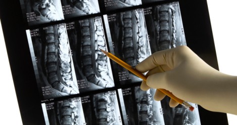
Scoliosis Associated with Friedreich’s Ataxia

The incidence of Friedreich’s Ataxia (FA) is approximately one in every 50,000. It is known that most symptoms associated with FA occur during the first decades of life. Many lose their ability to walk by age 25. Friedreich himself noted the presence of scoliosis along with other symptoms in 1875. Scoliosis is associated with FA and is often seen early on in the developmental process. A search of the literature finds that the prevalence of scoliosis among those with FA has been reported in the range of 63 to 100%1, 2, 3

There is some debate noted on whether FA should be classified as a neuromuscular scoliosis or idiopathic type. For the most part, scoliosis associated with FA is similar to other neuromuscular forms such as muscular dystrophy which features a progressive thoracolumbar curve and severe muscle weakness. Labelle et al (1986) concluded that scoliosis associated with FA behaved more like an idiopathic form as the curves were not necessarily rapidly progressive, were similar curve patterns, were not associated with muscle weakness, and age at onset was a key indicator of progression4. This was based on a retrospective review of 56 cases with an average of nine years of follow-up after the diagnosis was made. This is contrary to the findings of Milbrandt et al (2008) who felt the curve patterns were more in-line with neuromuscular type scoliosis.

The current literature does reflect bracing does not appear to halt progression of most curves that have been studied. A posterior spinal fusion has become the main treatment choice for surgical management of progressive curves associated with FA. Previously, hook and wire constructs were utilized but has fallen out of favor due to inability to maintain correction. Many of the curves are double major type with kyphotic deformity. Restricted lung disease associated has been reported as a secondary complication in cases with significant kyposcoliotic deformity. Cardiac co-morbidities are also associated with FA and must be considered when contemplating surgical management.

Despite known complications associated with FA, reported outcomes from posterior fusion surgery have demonstrated successful correction of the major deformity and fusion rates at an average of 3 years of follow-up.

1 Milbrandt TA, Kunes JR, Karol LA. Friedreich’s ataxia and scoliosis: the experience at two institutions. Journal of Pediatric Orthopedics. 2008 Mar; 28(2) 234-238.

2 Daher YH, Lonstein JE, Winter RB, Bradford DS. Spinal Deformities in patients with Friedreich ataxia: a review of 19 patients. Journal of Pediatric Orthopedics. 1985 Sep-Oct; 5(5): 553-557.

3 Cady RB, Bobechko WP. Incidence, natural history, and treatment of scoliosis in Friedreich’s ataxia. Journal of Pediatric Orthopedics. 1984 Nov; 4(6) 673-676.

4 Labelle H, Thome S, Duhaim M, Allard P. Natural history of scoliosis in Friedreich’s ataxia. JBJS. 1986 Apr; 68(4): 568-572.

Does Coffee Drinking Lower the Risk of Melanoma?

19th Feb 2015 Diseases, Medical News

A study published recently by researchers from the Yale School of Public Health and the Division of Cancer Epidemiology and Genetics at the National Institute of Health (NIH), reported the consumption of coffee was associated with a reduced risk of melanoma.

It is estimated that approximately one in five Americans will develop skin cancer.  Melanoma is currently the leading cause of skin cancer death in the U.S. with an increasing incidence.   Nearly 5000 Floridians are diagnosed with Melanoma annually.

Estimates of coffee consumption reveal that that Americans drink an average of 3.1 cups of coffee daily (NCA Drinking Trends Survey).  Furthermore, 83% of U.S. adults drink coffee.

The recent study looked at 447,357 non-Hispanic whites who were cancer free during at baseline.  They were followed for an average of 10.5 years. During the study period, 2904 developed melanoma.  Using statistical methods to determine risk, those who were in the group with the highest coffee consumption had the lowest risks of developing melanoma. Even after controlling for confounding variables including sex, age, BMI, alcohol intake, smoking status, and ultraviolet radiation exposure, the results held true.

Prior studies looking at the association between coffee consumption and melanoma have had mixed results. In a 2012, published data derived from the Nurses’ Health Study and Health Professionals Follow-up Study found that those women who consumed the most caffeine had an 18% lower risk of basal cell cancer (Song F, Qureshi AA, and Han J.  Cancer Research 2012 Jul 1; 72(13) 3282-3289).  However, caffeine intake was not associated with a lower risk of melanoma. Contrary to this, a prior case control study based out of Italy did show a protective effect of daily coffee consumption on the development of melanoma.

Studies have shown that the components of coffee may protect against oxidative stress and damage to DNA. There is belief that coffee may suppress Ultraviolet (UVB) induced skin cancers. Animal models have shown that caffeine helps to eliminate damaged skill cells before they have a chance to develop into tumors.

Caution should be exercised in considering these results as conclusive.   More research is needed to look at this association.   Differences in populations, possible measurement errors, and other confounding variables must be considered. Furthermore, while the lowest risk of developing melanoma in this study was associated with those who consumed the most coffee, there are known side effects of excessive coffee consumption.

Craiglist and HIV: Back to Basics

12th Feb 2015 Diseases, Medical News, Viruses

With the recent study released by the University of Minnesota linking Craigslist personals with an increase in reported HIV cases, it may be a good time to review this disease that has, in a way, fallen under the radar for many individuals. What is HIV? Where does it come from? How can you protect yourself? These are all questions that everyone should know how to answer.

What is HIV?

HIV is the initialism for human immunodeficiency virus. This virus is a type of retrovirus that causes AIDS, or acquired immunodeficiency syndrome. This disease causes progressive loss of immune system function, which can result in infections and cancers.

How does HIV damage the immune system?

HIV attacks certain immune system cells and directly or indirectly causes their death. When the number of these cells gets too low the individual loses cell-mediated immunity, which is a part of the immune response that does not use antibodies.  Without cell-mediated immunity opportunistic infections can take hold.

What are the symptoms of HIV?

Many people with HIV aren’t even aware they have the virus. Symptoms generally appear about a month or two after infection, but they can be so mild that they go unnoticed. These symptoms include: fever, fatigue, diarrhea, coughing, weight loss, shortness of breath, and swollen lymph nodes. The swollen lymph nodes are often the first signs of HIV infection. Some people describe it as “the worst flu ever.”

Without treatment HIV will progress and symptoms may include shaking chills, fever, night sweats, lesions of the tongue and mouth, blurred vision, and skin rashes. Over time cancers and opportunistic infections will develop and will be the cause of death if untreated.

How does someone contract HIV?

HIV can be spread through unprotected sexual intercourse (oral, anal, or vaginal), through blood transfusions (US hospitals screen for HIV to prevent this), sharing contaminated needles, and through pregnancy or breast-feeding. The virus is present in blood, semen, vaginal fluid, pre-ejaculate, and breast milk.

When does HIV become classified as AIDS?

The current standard for diagnosing AIDS is: a positive HIV test along with a CD4 cell count below 200, AND the presence of an AIDS-defining complication. This complication can be a disease like pneumocystosis, tuberculosis, toxoplasmosis, or many others.

How is HIV treated?

There is no cure for HIV or AIDS, but many drugs can be used to manage symptoms. Physicians usually prescribe multiple different types of medication to avoid developing HIV strains that are immune to single drugs. Treatment involves taking multiple pills at certain times every day for the rest of the patient’s life. These drugs can in turn cause their own side effects and if other health issues are present they may interfere with the individual’s ability to tolerate treatment.

Treatment can help a person life a normal life span, but without treatment life-expectancy is only 3 years.

How can I protect myself from HIV?

There is no vaccine available for HIV, but there are other methods you can use to protect yourself. The biggest way you can protect yourself is to use a new condom every time you have sexual intercourse (either vaginal or anal) and use a dental dam during oral sex. There is a drug called Truvada that MAY reduce the risk of sexually transmitted HIV if you are a high-risk individual (have unprotected sex, use IV drugs, are an uncircumcised male, or have another sexually transmitted disease.)

You should also use a new, clean needle every time if you are injecting drugs. There are needle-exchange programs available in many communities that you can use. Even better – seek help for your drug use.

If you are HIV-positive be sure to inform your sexual partners. If you become pregnant you need to speak to your doctor immediately about treatment to reduce the chance of passing the disease on to your baby.

MOXle Study: Evaluating the drug RTA 408 in the treatment of Friedreich’s Ataxia.

A clinical trial for a novel medication to treat Friedreich’s Ataxia was announced this past week. Reata Pharmaceuticals, a privately held biopharmaceutical company based out of Texas, announced the beginning of enrollments for a phase 2 and 3 clinical study titled “MOXle – RTA 408 Capsules in Patients with Friedreich’s Ataxia.” The study aims to further evaluate the safety, efficacy and pharmacodynamics of the treatment medication. A randomized, double blind, study to evaluate the maximum tolerated dose will be part one of the study. Part two will consist of splinting participants into two groups and giving two different dose levels of RTA 408. The second part will also be a randomized, placebo-controlled, double blinded study. This will be a multi-center study in which 52 patients are being sought.

Two clinical interests of the study will be peak workload during exercise as well as the modified Friedreich’s ataxia rating scale (FARS). The latter is a measurement tool which monitors proficiency of patients during activities. Additional biochemical endpoints will also be evaluated. Treatment with the study medication or placebo will be given to patient’s once daily for twelve weeks.

Nrf2 or Nuclear Factor 2, is a protein that regulates the expression of antioxidant proteins that protect against oxidative damage. Friedreich’s Ataxia is caused by defects in the gene for frataxin which is involved in the regulation of iron levels in the mitochondria. In pre-clinical studies, lower expressions of Nrf2 were correlated with frataxin deficiency and lower mitochondrial function. It is believed that RTA 408 can activate the Nrf2 pathways and improve mitochondrial function. Taken from animal models, RTA 408 showed the ability to improve overall cellular metabolism.

The study is expected to achieve final outcome measures by the spring of 2015 and study completion closer to June 2016.

http://www.marketwatch.com/story/reata-enrolls-first-patient-in-the-moxie-study-a-phase-23-study-examining-rta-408-in-friedreichs-ataxia-patients-2015-01-29

http://clinicaltrials.pharmaceutical-business-review.com/news/reata-enrolls-first-patient-in-phase-iiiii-moxie-study-of-rta-408-in-friedreichs-ataxia-patients-300115-4500885

Hospital-acquired Infections

03rd Feb 2015 Diseases, News and Events, Viruses

Hospital-acquired infections have once again found a place in the news. Thirty-two individuals are reported to have been infected by a number of different drug-resistant strains of bacteria through contaminated endoscopes.  Eleven of these patients have died, but due to their prior severe illnesses it is unclear whether or not the new infections played any role. The endoscopes in question are called duodenoscopes and are used to treat liver and pancreas illnesses. They are professionally sterilized to high standards between patients, which has unfortunately turned out to no longer be adequate.

The disinfection procedures that the manufacturers recommended were approved by the U.S. Food and Drug Administration (FDA), but after the hospital was able to identify the sterilized scopes as the source of infection they had to switch to a method that exceeded the national standards. The FDA is working with the endoscope suppliers and medical centers to develop new solutions, but this is not the first time scopes have been identified as a source of nosocomial infections. Pittsburgh in 2012 and Chicago in 2014 both saw hospital-acquired infections due to contaminated endoscopes, but luckily there were no fatalities.

The most well-known hospital-acquired infection is Methicillin-resistant Staphylococcus aureus, or MRSA. MRSA originated in the hospital setting, but has expanded to locker rooms, livestock, prisons, military barracks, and homeless shelters. Since many of the individuals infected have weakened immune systems they are already at a greater risk of nosocomial infections. In addition, MRSA’s resistance to certain antibiotics (like penicillins and cephalosporins) make it very difficult to treat.

One of the drug resistant strains of bacteria responsible for the infections in Seattle include Carbapenem-resistant enterobacteriaceae (CRE). Two examples of CRE include Klebsiella species and Escherichia coli (E. coli). While these are normal human gut bacteria, they can cause infection in those undergoing invasive treatments due to illness (like the endoscopes) or those that are taking long courses of antibiotics. Many strains of CRE have become resistant to most of the available antibiotics, making them especially deadly.

How can you protect yourself from hospital-acquired infections, like MRSA and CRE? Most of the prevention is done by the hospital – sterilizing equipment, using appropriate isolation procedures, washing hands, wearing gloves, sanitizing surfaces, wearing aprons while treating patients, and even using antimicrobial surfaces like copper bedrails. For patients that want to protect themselves there is a list of questions you can ask your healthcare provider that is provided by the Centers for Disease Control and Prevention: http://www.patientcarelink.org/uploadDocs/1/Massachusetts-Consumer-HAI-Basics.pdf

The ‘Kissing Bug’ Disease: What You Need To Know

17th Nov 2014 Diseases, Medical News

Chagas disease, also known as the ‘kissing bug’ disease, is an infection that is affecting more and more Americans every year. Since this disease has primarily been found in places like Mexico as well as South and Central America, many people in the United States, including physicians, are unfamiliar with it. However, the last few years have seen cases turning up in the United States. Most people are believed to have been infected abroad, but recently more of these infections have been contracted locally.

The kissing bug (also known as the triatomine bug, reduviid bug, assassin bug, cone-nosed bug, or blood sucker) includes many different species that can all carry the parasite Trypanosoma cruzi that causes Chagas disease. These insects can be found both indoors and outdoors, particularly in the cracks and holes found in substandard housing. They are found across the southern two-thirds of the United States, but the parasite is generally found in the triatomine bugs from Latin America. There are other bugs that resemble this insect (like the assassin bug or the wheel bug), so if one is found it can be taken to an entomologist for positive identification.

Fortunately, contracting Chagas disease from kissing bugs is rather difficult – an individual needs to contact the insect’s feces through a wound (like the bite wound that the insect makes, often on the face) or a mucous membrane (like the mouth or the eyes.) Some people are allergic to the insect’s saliva, but this does not mean that they have contracted the disease. Signs of an allergic reaction include redness, itching, swelling, and welts or hives. In rarer cases this reaction can cause anaphylactic shock.

In the event that someone does contract Chagas disease there may be a mild swelling at the site where the parasite entered the body – this is known as a chagoma. Romaña’s sign is swelling around the eye if the eye was the parasite’s inoculation point. The chagoma or Romaña’s sign both last longer than an allergic reaction and are not nearly as itchy. Other than the potential swelling and fever, there are very few early signs for Chagas disease. In rare cases there may be severe inflammation around the heart muscle, brain, and lining of the brain.

If a person remains untreated they can enter a chronic phase of disease where very few or no parasites are found in their blood, making diagnosis difficult. Many people will remain symptom free and never know they have the disease, but 20-30% can develop symptoms. These symptoms include heart rhythm abnormalities that can cause sudden death, a dilated heart that doesn’t pump well, and a dilated esophagus or colon that makes it difficult to eat or to pass stool.

Chagas disease can be spread through blood transfusions, organ transplants, and congenitally from mother to child. Many people have been notified that they test positive for the parasite after donating blood, despite not being aware that they had it. Two drugs can be used to treat the disease if it is caught early enough – nifurtimox and benznidazole. Due to the more “exotic” nature of this disease many physicians aren’t familiar with it. If you are concerned that you may have been exposed to or contracted Chagas disease you can talk to your physician or seek one familiar with the disease here: http://www.astmh.org/source/ClinicalDirectory/


Dr. Susan Perlman and Stephanie Magness on American Health Journal Nov 7

Dr. Susan Perlman appeared on American Health Journal this Friday to discuss Friedreich’s Ataxia (FA) along with one of her patients, Stephanie Magness. They educated viewers on the clinical aspects of the disease as well as daily life and clinical studies.

Dr. Perlman is a professor in the Department of Neurology at the David Geffen School of Medicine at UCLA and physician specializing in ataxia, Huntington’s Disease, and neurogenetics. She is the Director of the Ataxia and Neurogenetics Program and the Post-Polio Program at UCLA and has been a primary investigator for many Friedreich’s Ataxia trials over the years.

In this program, Dr. Perlman reviews some of the basic facts of the disease – it is a recessive neurogenetic disorder caused by mutations in both copies of the gene that controls the production of frataxin, it is a disease of childhood, and the symptoms start with balance and coordination difficulties. It begins in the legs and progresses to the hands, speech, vision, and even hearing. The patients may also develop cardiac symptoms, which include hypertrophic cardiomyopathy, heart rhythm problems, and even heart failure. Diabetes is another potential symptom and can cause affected individuals to require insulin.

Stephanie Magness is currently one of Dr. Perlman’s patients and, like all too many people with FA, she was not diagnosed correctly for the first few years of her symptoms. Friedreich’s Ataxia is often misdiagnosed as another recessive childhood neurogenetic disease like Charcot-Marie-Tooth disease, cerebral palsy, and idiopathic peripheral neuropathy. She was eventually diagnosed correctly via gene testing and is now able to manage her symptoms accordingly. Stephanie has also taken part in a natural history study as well as a clinical drug trial – possibly the first approved drug for FA.

According to Stephanie one of the most difficult aspects of the disease is its progressive nature. Even if a patient is able to handle their symptoms well currently they know that it may all change. Her advice was to focus on what you can do instead of things that you obviously cannot and find new things to do that you can excel in.

Now that drug companies have begun to take an interest in Friedreich’s Ataxia and to see it as an important area scientifically there is hope that a drug will be approved to treat FA in the not-too-distant future. Watch the program on Vimeo here: http://vimeo.com/110841193 (the section about Friedreich’s Ataxia starts at 15:39)


Saint Louis Encephalitis

31st Oct 2014 Diseases, Medical News, Viruses

Over the summer there have been mentions of Saint Louis encephalitis virus (SLEV) in regards to Pinellas County. This virus is a flavivirus – a group that includes West Nile – and has the potential to cause severe illness or death in older adults. The name of this disease comes from a particularly extensive outbreak that occurred in St. Louis, Missouri and the surrounding St. Louis County in 1933. Due to the high number of cases NIH was able to investigate and isolate this previously unknown virus. It is thought to have originated in northern Mexico and have been carried north by birds. Pinellas County Mosquito Control uses sentinel chickens to monitor for Saint Louis encephalitis and other arboviruses, since birds are usually the source of infection for mosquitos. Blood is drawn from the chickens weekly and tested for antibodies to the viruses of concern. Fortunately for the chickens they do not develop any symptoms, although once they test positive they have to be removed from the program since they will always carry the antibodies. Sentinel chickens from Pinellas County have tested positive for Saint Louis encephalitis in late July as well as August and into September. Regions with confirmed SLEV include Cross Bayou, Walsingham Park, Sawgrass Lake Park, and Lake Maggiore. While it is more common during hot and humid months, Saint Louis encephalitis can occur year round. Most individuals that are infected have no symptoms, but in rare cases it can manifest with such signs as fever, headache, nausea, vomiting, and fatigue. Severe infections can result in high fever, neck stiffness, disorientation, confusion, tremors, coma, and sometimes convulsion, spastic paralysis, and death. Older adults are at a greater risk for fatal disease. Since there is no cure for Saint Louis encephalitis the only treatment available is supportive care. If you or anyone you know shows symptoms of this disease be sure to talk to your health care provider for proper diagnosis. Since this virus is spread by mosquitos you should take the appropriate precautions. Wear insect repellent and protective clothing and be sure to avoid areas with high mosquito populations. Drain standing water, change pets’ water dishes and birdbaths regularly, and maintain your pool’s water balance to prevent mosquitos from breeding. If you use rain barrels be sure to cover them with fine mesh. Ornamental ponds can be stocked with fish that eat the mosquito larvae and any ornamental bromeliads should be flushed with fresh water regularly and treated with a larvicide. Mosquito Control field technicians can respond to concerns by use of the Mosquito Control Request form found on the Pinellas County website. http://www.pinellascounty.org/forms/mosquito_form.htm

Beware of Poisonous Caterpillar in Florida

25th Sep 2014 Diseases, Medical News

Recently there have been countless news stories about a spectacular caterpillar – the puss caterpillar. Children from Florida to Virginia have been stung and the excruciating pain has been landing them in the hospital. Some of these children had actively sought out to pet the caterpillar due to its “cute” fuzzy appearance. In fact, the caterpillar gets its name due to the fact that it resembles a “fluffy house cat” or even a “tiny Persian cat.” Other children came into contact when the insect fell from a tree and landed on them.

These caterpillars are generally an inch long at the largest and are covered in a thick coat of fur that can range from greyish-white to orangey-brown to charcoal. In addition to the fur their body tapers at the end, which can give it the appearance of having a tail. The fluffy hairs that cover the puss caterpillar hide a series of tiny, venomous spines that can stick in your skin.

The feeling is said to be similar to a bee sting, except worse, and immediately starts increasing to the point where it can even “make your bones hurt.” The degree of pain depends on where you’re stung, but it has been reported to take as many as 12 hours to begin to subside. On top of the intense pain, contact with the caterpillar’s bristles can cause fever, vomiting, convulsions, rashes, swelling, abdominal distress, blisters, and even chest pain, numbness, and difficulty breathing. However, each individual reacts to a different extent – some require medical treatment and other report only a mild itching or burning.

It may seem like more and more people are being stung recently, which is partly true. Like many other insect species, the puss caterpillar populations go through a “boom-and-bust” cycle. This means that the population can increase or decrease quite dramatically as environmental conditions change. Right now these conditions appear to be ideal for the caterpillars so the population has been on the rise.

The Poison Center of Tampa recommends that you use cellophane tape over the affected area and remove repeatedly to strip the spines from your skin. In addition, you can apply ice packs to reduce the stinging sensation and follow it up with applying a paste made of baking soda and water. Oral antihistamines, calamine lotion, hydrocortisone cream, and juice from the stems of comfrey plants can also be used to treat symptoms. It is recommended to watch for the caterpillars around trees like oak, elm, and citrus. They even tend to be found around common garden plants like rose bushes or ivy. Additionally, teach children what they look like and why they need to avoid touching them since the puss caterpillar’s fluff-ball appearance can be quite appealing.

Puccio Study FA

19th Sep 2014 Diseases, Friedreich's Ataxia

A recent breakthrough in Friedreich’s ataxia (FA) research gives hope that gene therapy can prove be an effective treatment for the disease. A team led by Hélène Puccio, Ph.D., has demonstrated that gene therapy can be used to reverse heart damage caused by FA and correct mitochondrial metabolism. Dr. Puccio works as the head of a research team at the Institute of Genetics and Molecular and Cellular Biology located at the University of Strasbourg, France, as well as research director at the Insititute de Santé et de la Recherche Médicale (INSERM.) She and her team have concentrated their research on the pathophysiological mechanisms involved in recessive ataxias, like FA, in order to develop new animal and cell models.

Friedreich’s ataxia is rare, but one of the more common recessive ataxias – found in 1 in every 50,000 births. It is caused by a mutation in the frataxin (FXN) gene, which in turn causes a decrease in the amount of frataxin produced. The lack of frataxin disrupts the mitochondria that are responsible for the cell’s energy production, and, unfortunately, heart tissue is particularly vulnerable.

While FA causes progressive neurodegeneration that starts as impaired balance and coordination, it also impairs heart function and can contribute to diabetes. Most patients require a wheelchair after about 10-20 years of disease progression, but complications with the heart lead to death in more than half of individuals, and often before the age of 35.

Dr. Puccio’s team used a harmless viral vector to insert a normal copy of the problematic gene in the heart cells of FA mouse models. Since these mice show the same heart symptoms as human patients suffering from the disease the results are especially promising. The virus used was an adeno-associated virus (AAV) since it is known to effectively and efficiently target heart cells and express therapeutic diseases. This virus was modified to render it harmless and only capable of inserting the normal FXN gene copy into the cells.

One single intravenous injection was not only able to prevent the development of heart disease in the mice, but it quickly and completely reversed the heart damage in advanced cases. Within three weeks the mice appeared to have completely restored heart and mitochondrial function and their heart tissue appeared similar to healthy mice.

Since this is the first time that gene therapy has produced such a fast and complete remission of heart disease in an animal model work is rapidly being done to begin clinical studies. There is also hope that this technique can be used to prevent or even correct damage to the spinal cord and cerebellum.